
688
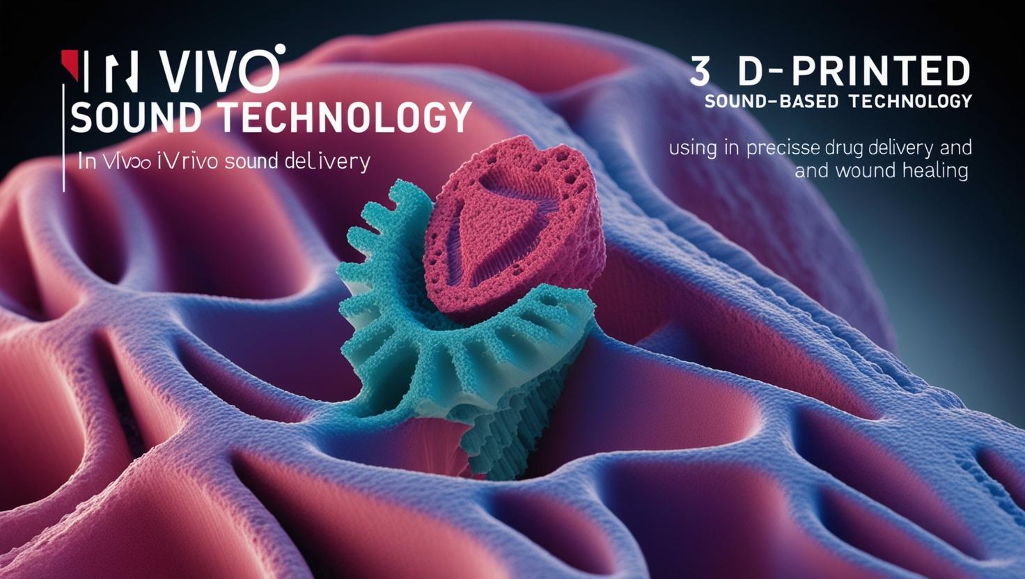
In Vivo 3D Printing Using Sound: Caltech Researchers Pioneer DISP Platform for Precision Deep Tissue Therapeutics
A research team led by the California Institute of Technology has unveiled a transformative innovation in biomedical engineering: Deep Tissue In Vivo Sound Printing (DISP). This novel platform enables high-resolution, targeted 3D printing of biopolymers and therapeutic payloads deep within living tissue, overcoming previous limitations posed by conventional light-based printing approaches. The technique promises to revolutionize drug delivery, tissue regeneration, and implantable bioelectronics, marking a significant leap forward in non-invasive therapeutic interventions.
The Challenge of Deep Tissue Bioprinting
Traditional in vivo polymerization techniques rely heavily on infrared (IR) light to initiate the crosslinking of monomers into functional polymers. However, IR light suffers from limited tissue penetration, restricting its effective depth to superficial layers just beneath the skin. This bottleneck has hindered the development of truly internal therapeutic structures, such as drug reservoirs or bioadhesive patches, within complex anatomical environments.
To address this challenge, Professor Wei Gao, a leader in medical engineering at Caltech, and his multidisciplinary team turned to a more penetrative and widely used biomedical modality: ultrasound. Their goal was to harness ultrasound not just for imaging or ablation, but as a localized trigger for precision bioprinting within living organisms.
Engineering the DISP Platform
At the heart of the DISP platform lies an ingeniously designed composite bioink, consisting of the following components:
Low-temperature-sensitive liposomes, pre-loaded with a chemical crosslinking agent.
Polymer monomers that can be crosslinked into gels or solids.
Imaging contrast agents, specifically bacterial gas vesicles, to monitor in situ polymerization.
Cargo payloads such as therapeutic drugs, cells, or conductive materials (e.g., silver or carbon nanotubes).
This composite bioink is injected into the target site within the body. To activate the printing process, the researchers use focused ultrasound to raise the local tissue temperature by approximately 5°C. This minor but precise thermal increase is sufficient to destabilize the liposomes, causing them to rupture and release the crosslinking agents.
Upon release, these agents initiate localized polymerization, effectively “printing” a three-dimensional structure in vivo. Notably, the incorporation of gas vesicles, which respond to the chemical environment during gelation, provides a real-time ultrasound imaging readout that reveals when and where polymerization has occurred.
Applications in Drug Delivery and Tissue Engineering
To demonstrate the therapeutic potential of the DISP platform, Gao’s team conducted preclinical experiments on mouse models of bladder cancer. When doxorubicin, a potent chemotherapy drug, was embedded in the DISP-formulated hydrogel and printed near tumor sites, the localized drug delivery achieved significantly higher tumor cell death compared to traditional drug injection methods.
Beyond chemotherapy, the team successfully printed:
Bioadhesive polymers for sealing internal wounds,
Hydrogels embedded with conductive materials for bioelectronic applications like ECG signal monitoring.
These versatile demonstrations suggest that the DISP platform is modular and adaptable to a wide array of biomedical contexts, including regenerative medicine, soft robotics, and implantable biosensors.
Advancing Toward Autonomous, AI-Guided Bioprinting
Looking forward, the team envisions integrating machine learning algorithms to enhance the spatial precision and automation of DISP. With the potential to compensate for tissue movement, such as that seen in beating hearts, AI-guided DISP could enable real-time, autonomous 3D bioprinting in dynamic biological environments.
According to Gao, “In the future, with the help of AI, we aim to develop systems capable of autonomously initiating high-precision printing inside moving organs such as a beating heart—enabling targeted therapeutic interventions that were previously inconceivable.”
Future Directions and Clinical Translation
While current validation is limited to small-animal models, the researchers are actively working to scale the technology for larger organisms, including human trials. Key focus areas include:
Optimization of bioink formulations for different therapeutic applications,
Integration with robotic and laparoscopic systems for minimally invasive deployment,
Regulatory pathways and biocompatibility validation for human use.
The study, titled “Imaging-Guided Deep Tissue In Vivo Sound Printing”, appears in the journal Science and represents a collaborative effort between Caltech and the University of Utah.
Reference:
Davoodi, E., et al. (2025). Imaging-guided deep tissue in vivo sound printing. Science. DOI: 10.1126/science.adt0293
Credit:– phys.org , or science.org
You Might Be Interested In
- The Complete Guide to Nanotechnology and Nanoscience Education: Top Colleges, Costs, Careers, and Specializations
- Top 10 Nanotechnology Innovations Set to Revolutionize 2025
- Career Paths in Nanotechnology: A Comprehensive Research-Style Overview
- The Importance of Scanning Electron Microscopy (SEM) in Modern Science and Technology