
627
Quantum dots (QDs) have emerged as a transformative nanotechnology platform in oncology, addressing critical limitations of conventional cancer treatments such as nonspecific toxicity and poor therapeutic monitoring. This review comprehensively examines the role of QDs in targeted drug delivery, leveraging their tunable optical properties, fluorescence stability, and quantum confinement effects. The physicochemical properties of QDs, including their high surface-to-volume ratio and photoluminescence, enable their integration into theranostic systems that combine diagnostics and therapy. Recent advancements in QD functionalization—such as biodegradable nanostructures, ligand-mediated targeting, and hybrid systems with chemotherapeutic, immunotherapeutic, or radiotherapeutic agents—are critically analyzed. Preclinical and emerging clinical data highlight the translational potential of QD-based platforms, focusing on their imaging resolution, pharmacokinetics, and biocompatibility. The review also explores QD-enabled theranostics for simultaneous tumor visualization and controlled drug release, while addressing challenges like long-term toxicity, scalability, and regulatory hurdles. By presenting QDs as a cornerstone for individualized, real-time cancer treatment, this study underscores the need for interdisciplinary collaboration to bridge the gap between bench and bedside.
1. Introduction
1.1 Background and Rationale
Cancer remains a leading global health challenge, characterized by high morbidity and mortality rates due to its systemic nature and complex interactions with the immune system. Traditional treatments, including chemotherapy, radiotherapy, surgery, and immunotherapy, often face significant limitations such as multidrug resistance (MDR), nonspecific toxicity, and limited bioavailability, which compromise their efficacy. The tumor microenvironment (TME) further complicates therapeutic outcomes by hindering immune responses outside its boundaries, necessitating a deeper understanding of systemic immunology for effective interventions.
The advent of nanotechnology has introduced nano-sized drug delivery systems (NDDSs) as a promising avenue to overcome these challenges. NDDSs, including liposomes, nanoparticles, and dendrimers, offer targeted drug delivery, deep tissue penetration, and reduced systemic toxicity. Among these, quantum dots (QDs)—semiconductor nanostructures with unique optical and electronic properties—stand out as versatile platforms for cancer theranostics. QDs exhibit tunable fluorescence, high photostability, and quantum confinement effects, making them ideal for simultaneous imaging and drug delivery. This review explores how QDs can revolutionize targeted drug delivery in oncology, addressing gaps in current therapeutic approaches and paving the way for personalized medicine.
1.2 Aim and Scope of the Study
This study aims to provide a comprehensive analysis of QD nanotechnology in advancing targeted drug delivery for cancer therapy. It focuses on the physicochemical properties of QDs, their mechanisms of action, and their integration with other therapeutic modalities. The scope includes an evaluation of their theranostic potential, preclinical and clinical advancements, and the challenges hindering their clinical adoption. The potential benefits of QD-based systems in oncology include reduced side effects through precise targeting, an enhanced therapeutic index, and cost-effectiveness by combining diagnostics and therapy into a single platform.
2. Quantum Dots: Fundamental Properties and Biomedical Potential
2.1 Physicochemical Properties of Quantum Dots
Quantum dots are semiconductor nanostructures, typically ranging from 2 to 10 nanometers in diameter, with properties governed by quantum mechanics. Their small size leads to quantum confinement effects, where excitons (electron-hole pairs) are confined in three dimensions, resulting in tunable optical properties. Smaller QDs (2–3 nm) emit shorter wavelengths (e.g., blue or green), while larger QDs (5–6 nm) emit longer wavelengths (e.g., orange or red). The composition of QDs, such as cadmium selenide (CdSe), zinc sulfide (ZnS), or carbon-based forms, further influences their emission spectra.
Graphene quantum dots (GQDs), a subset of carbon-based QDs, exhibit exceptional characteristics, including high photoluminescence, biocompatibility, and a large surface-to-volume ratio. Their honeycomb-like lattice (sp2 hybridization) and zero-dimensional structure make them highly dispersible and stable in physiological environments. Unlike graphene oxide (GO), which requires additional functionalization for stability, GQDs are inherently hydrophilic, facilitating the delivery of partially water-soluble drugs. The adjustable bandgap of GQDs, influenced by their size and edge effects, enhances their optoelectronic properties, making them suitable for bioimaging and drug delivery applications.
2.2 Biomedical Relevance
QDs offer significant advantages in biomedical applications due to their optical and functional properties. Their high quantum yield, narrow emission spectra, and resistance to photobleaching make them superior to organic dyes for imaging purposes. GQDs, in particular, combine the benefits of graphene (large surface area, biocompatibility) with QD-specific properties (fluorescence, quantum confinement), enabling efficient drug loading and real-time tracking without additional marker dyes. However, concerns about genotoxicity—stemming from heavy metal content (e.g., cadmium, lead) or reactive oxygen species generation—require further investigation to ensure safe clinical use.
3. Mechanism of QD-Mediated Targeted Drug Delivery
3.1 Principles of Targeted Delivery Using QDs
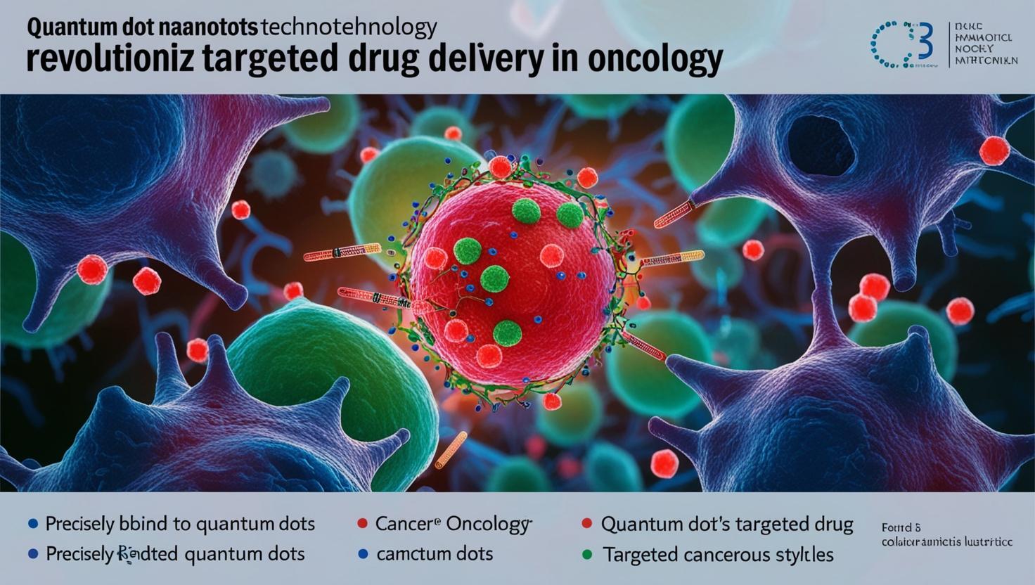
QDs enable both passive and active targeting strategies for drug delivery in oncology. Passive targeting exploits the enhanced permeability and retention (EPR) effect, where the leaky vasculature of tumors allows nanoparticles to accumulate preferentially. Small-sized QDs (less than 10 nm) exhibit improved tumor penetration and longer circulation half-lives, enhancing drug delivery efficiency. Active targeting, achieved through surface functionalization with ligands (e.g., antibodies, peptides, or aptamers), increases specificity by binding to overexpressed receptors on cancer cells, such as epidermal growth factor receptor (EGFR) or folate receptors.
A notable example is the use of sulfonic-graphene quantum dots (sulfonic-GQDs) for nuclear targeting in tumor cells. The increased interstitial fluid pressure (IFP) in tumors facilitates the selective entry of sulfonic-GQDs into cancer cell plasma membranes, while their negative charge prevents uptake by healthy cells. Molecular dynamics simulations demonstrate that these GQDs can cross the plasma and nuclear membranes, enabling precise drug delivery and tumor-specific imaging.
3.2 Integration with Other Therapeutic Modalities
QDs can be integrated with photodynamic therapy (PDT) and photothermal therapy (PTT) to enhance therapeutic outcomes. In PDT, QDs act as photosensitizers, generating singlet oxygen upon light activation to kill cancer cells. In PTT, QDs convert near-infrared (NIR) light into heat, inducing thermal ablation of tumors. Förster resonance energy transfer (FRET) mechanisms further improve the efficiency of these therapies by enabling energy transfer between QDs and photosensitizers. Hybrid systems, such as QD-liposome complexes (QD-L), reduce cytotoxicity while maintaining imaging and drug delivery capabilities, offering a multifunctional platform for cancer treatment.
4. Innovations in Quantum Dot Design for Oncology
4.1 Engineering Quantum Dots for Enhanced Efficacy
Recent advancements in QD synthesis focus on improving their fluorescence and biocompatibility. Surface passivation with polymers like polyethylene glycol (PEG) enhances the solubility and fluorescence of GQDs, as seen in GQDs-PEG formulations. Doping with heteroatoms (e.g., nitrogen, phosphorus) or chemical treatments (e.g., acid/base modification) allows for fine-tuning of optoelectronic properties, optimizing QDs for specific applications. These engineering strategies improve drug loading, stability, and targeting efficiency, making QDs more effective in oncology.
4.2 Biodegradable Quantum Dots
Traditional QDs containing heavy metals (e.g., CdSe, PbS) pose long-term toxicity risks due to bioaccumulation. Biodegradable QDs address these concerns by using safer materials, such as silica, carbon, or biodegradable polymers like poly(lactic-co-glycolic acid) (PLGA). Carbon quantum dots (CQDs) exhibit excellent biocompatibility and degrade into non-toxic byproducts, reducing systemic toxicity and facilitating clearance from the body. Surface modifications with hydrophilic coatings further enhance biodistribution and degradation, improving the safety profile of QDs for clinical applications.
5. Theranostic Applications of Quantum Dots in Oncology
5.1 Dual Functionality: Imaging and Drug Delivery
QDs enable theranostic applications by combining imaging and drug delivery in a single platform. Their fluorescence properties allow for high-resolution imaging of tumors, while their large surface area facilitates drug loading. For example, PEG-GO-Fe3O4 nanocomposites deliver melittin (MEL) to cancer cells while enabling real-time monitoring of drug release. Functionalized graphene-based materials, conjugated with targeting ligands like folic acid (FA) or monoclonal antibodies, enhance specificity for tumor cells, supporting applications in smart drug delivery, PTT, and PDT.
5.2 Imaging Modalities for Monitoring QD Distribution In Vivo
QDs support multiple imaging modalities for tracking their distribution in vivo, including fluorescence imaging, NIR imaging, magnetic resonance imaging (MRI), photoacoustic imaging (PAI), positron emission tomography (PET), computed tomography (CT), and single-photon emission computed tomography (SPECT). NIR fluorescence imaging, in particular, offers deep tissue penetration (up to several centimeters) and low autofluorescence, making it ideal for tumor imaging and biodistribution studies. These modalities enable comprehensive monitoring of QD behavior, therapeutic efficacy, and clearance.
5.3 Clinical Potential of Quantum Dots
QDs have demonstrated clinical potential in various cancer types. In ovarian cancer, QD probes detect the CA125 antigen with higher sensitivity than traditional organic dyes, enabling early diagnosis. In liver cancer, QD-Anti-AFP probes facilitate spectroscopic imaging of tumors, tracking progression and metastasis. In lung cancer, QD-based immunohistochemistry (QD-IHC) outperforms traditional IHC in detecting caveolin-1 and PCNA antigens, offering improved sensitivity and photostability for tissue microarrays.
6. Real-Time Monitoring and Personalized Medicine
6.1 Real-Time Monitoring
QDs, particularly CQDs, enable real-time monitoring of cancer progression and treatment response due to their high photostability and tunable fluorescence. Functionalized CQDs can bind to tumor-specific biomarkers, providing dynamic imaging of tumor growth, angiogenesis, and metastasis. They also support multiplexed imaging, allowing simultaneous monitoring of multiple biomarkers, and can be integrated with advanced techniques like photoacoustic imaging for deeper tissue visualization. These capabilities enhance early detection, therapeutic evaluation, and disease relapse monitoring.
6.2 Personalized Medicine
QDs advance personalized medicine by enabling targeted drug delivery, theranostic integration, and multi-modal therapy tailored to individual patients. They can deliver drugs to specific tumor sites, monitor treatment response in real-time, and detect genetic mutations (e.g., KRAS, BRAF) for precision diagnosis. By combining diagnostics and therapeutics, QDs facilitate dynamic therapy adjustments, reduce off-target effects, and improve patient outcomes through customized treatment plans.
7. Preclinical and Clinical Advancements
7.1 Preclinical Studies: Efficacy and Safety
Preclinical studies demonstrate the efficacy of QDs in imaging and targeting various cancer types, including melanoma, breast, and lung cancers. For example, anti-IGF1R QDs effectively image breast cancer cells (MCF-7), while nitrogen-phosphorus-doped CQDs induce autophagy and cell death in melanoma cells (B16F10). In vivo studies using ternary copper indium selenide/zinc sulfide QDs show promise for NIR bioimaging and imaging-guided surgery in head and neck squamous cell carcinoma. Safety assessments highlight the need for biodegradable QDs to mitigate toxicity concerns.
7.2 Clinical Trials and Regulatory Pathway
While most QD research remains preclinical, emerging clinical trials focus on their safety and efficacy as diagnostic and theranostic agents. Regulatory pathways involve preclinical safety testing, Investigational New Drug (IND) or Investigational Device Exemption (IDE) applications, and phased clinical trials. The U.S. FDA, European Medicines Agency (EMA), and International Council for Harmonisation (ICH) guidelines emphasize quality by design (QbD), long-term safety data, and good manufacturing practices (GMP). Challenges include scalability, standardization, and ethical concerns, necessitating robust regulatory oversight.
8. Discussion
8.1 Challenges and Future Directions
Despite their promise, QDs face challenges in clinical translation. Toxicity from heavy metals (e.g., cadmium, lead) raises concerns about bioaccumulation and genotoxicity, necessitating the development of low-toxicity alternatives like zinc-based or carbon-based QDs. Nonspecific organ uptake by the reticuloendothelial system (RES) and short circulation half-lives can be addressed through surface modifications with PEG or charge optimization. Standardization of QD synthesis and long-term safety studies are critical for ensuring reproducibility and regulatory approval.
8.2 Prospects for the Future
Future advancements in QD technology will focus on biodegradable, NIR-emitting formulations with enhanced safety profiles. Interdisciplinary collaboration between materials scientists, oncologists, and regulatory experts will accelerate the translation of QD-based theranostics into clinical practice, offering new hope for personalized and effective cancer treatments.
9. Conclusion
Quantum dots represent a paradigm-shifting nanotechnology for targeted drug delivery in oncology, leveraging their unique optical properties and functional versatility. Their ability to enable precise targeting, real-time monitoring, and theranostic applications positions them as a cornerstone for personalized medicine. While challenges such as toxicity, scalability, and regulatory hurdles remain, ongoing innovations in biodegradable QD design and preclinical/clinical research are paving the way for their integration into cancer care. Collaborative efforts across disciplines are essential to fully realize the potential of QDs in transforming oncology.
References
The original content is derived from the www.sciencedirect.com
You Might Be Interested In
- Postdoctoral Research Opportunity in Image Processing for Bioimaging at TU Delft
- MIT Physicists Capture First Real-Space Images of Free-Interacting Atoms
- Top 10 Nanotechnology Institutes in the World: Leading the Future of Nano-Innovation
- Nanorobots in Biomedicine and Beyond: The Nano-Scale Agents Redefining Precision Therapeutics and Technological Innovation